Right Aortic Arch and Arterial Flow Steal: Case Report
Article Sidebar
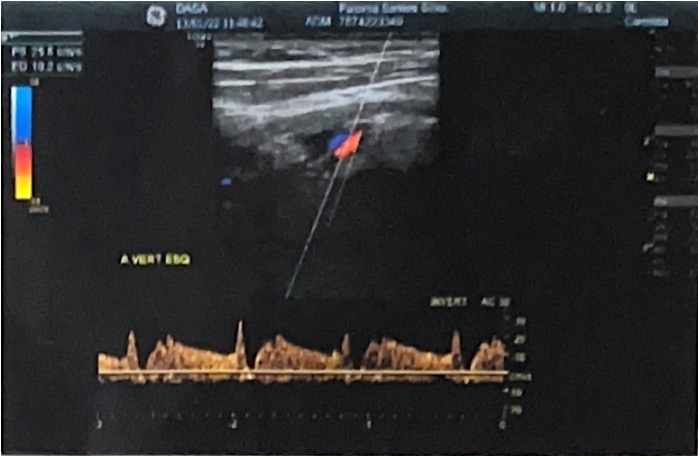
Main Article Content
Abstract
Article Details

This work is licensed under a Creative Commons Attribution 4.0 International License.
Authors retain the copyright of their articles and grant the journal the right of first publication under the Creative Commons Attribution (CC BY) license, which allows others to share and adapt the work with proper attribution.
References
Triantafyllou G, Melissanidis S, Vlychou M, Tsakotos G, Pantazis N, Vassiou K, et al. Right-Sided Aortic Arch: A Computed Tomography Angiography Investigation, A Systematic Review with Meta-Analysis. Journal of Clinical Medicine. 2024 May 25;13(11):3105.
Türkvatan A, Büyükbayraktar FG, Ölçer T, Cumhur T. Congenital Anomalies of the Aortic Arch: Evaluation with the Use of Multidetector Computed Tomography. Korean Journal of Radiology. 2009;10(2):176.
Stojanovska J, Cascade PN, Chong S, Quint LE, Sundaram B. Embryology and Imaging Review of Aortic Arch Anomalies. Journal of Thoracic Imaging. 2012 Mar;27(2):73–84.
Açar G, Çiçekcibaşı AE, Uysal E, Koplay M. Anatomical variations of the aortic arch branching pattern using CT angio-graphy: a proposal for a different morphological classification with clinical relevance. Anatomical Science International. 2021 Sep 10;97(1):65–78.
Hanneman K, Newman B, Chan F. Congenital Variants and Anomalies of the Aortic Arch. RadioGraphics. 2017 Jan;37(1):32–51.
Backer CL, Mavroudis C. Congenital Heart Surgery Nomenclature and Database Project: patent ductus arteriosus, coarcta-tion of the aorta, interrupted aortic arch. The Annals of Thoracic Surgery. 2000 Mar;69(3):298–307.
Ramos-Duran L, Nance JW, U. Joseph Schoepf, Henzler T, Apfaltrer P, Hlavacek AM. Developmental Aortic Arch Anomalies in Infants and Children Assessed With CT Angiography. 2012 May 1;198(5):W466–74.
Hanneman K, Newman B, Chan F. Congenital Variants and Anomalies of the Aortic Arch. RadioGraphics. 2017 Jan;37(1):32–51.
Popieluszko P., Henry B.M., Sanna B., Hsieh W.C., Saganiak K., Pękala P.A., Walocha J.A., Tomaszewski K.A. A Systematic Review and Meta-Analysis of Variations in Branching Patterns of the Adult Aortic Arch. J. Vasc. Surg. 2018;68:298–306.e10. doi: 10.1016/j.jvs.2017.06.097.
Polguj M, Chrzanowski Ł, Kasprzak JD, Stefanczyk L, Topol M, Majos A. The aberrant right subclavian artery (arteria luso-ria) – the morphological and clinical aspects of one of the most important variations: a systematic study of 141 reports. Sci World J. 2014;2014:292734.
Arazińska A, Polguj M, Szymczyk K, Kaczmarska M, Trębiński Ł, Stefańczyk L. Right aortic arch analysis – anatomical variant or serious vascular defect? BMC Cardiovasc Disord. 2017;17:102. doi:10.1186/s12872-017-0536-z.
Law M.A., Mohan J. Right Aortic Arches. StatPearls Publishing; Treasure Island, FL, USA: 2024. https://www.ncbi.nlm.nih.gov/books/NBK431104/.
Backer CL, Mavroudis C. Ann Thorac Surg. 2000;69(Suppl 4):S298–S307.
Tsiouris C., Lazaridis N., Piagkou M., Duparc F., Antonopoulos I., Antonitsis P., Natsis K. The Left-Sided Aortic Arch Va-riants: Prevalence Meta-Analysis of Imaging Studies. Surg. Radiol. Anat. 2022;44:673–688. doi: 10.1007/s00276-022-02945-4.